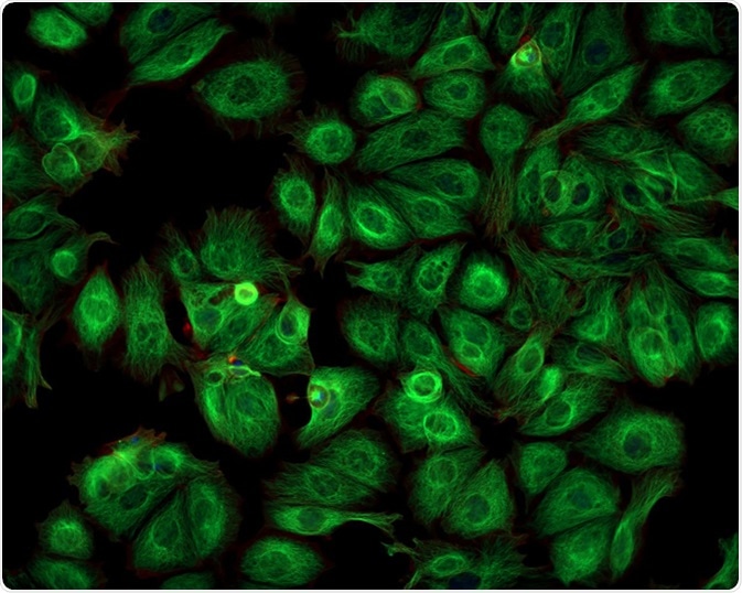
After the discovery of green fluorescent protein (GFP) in 1961, fluorescent proteins have been fused to various proteins and enzymes in order to visualize them. Fluorescent proteins ushered a new era in the field of cell biology, enabling live visualization of molecules and processes inside the cell.
The Discovery of Green Fluorescent Protein (GFP)
Two scientists, Osamu Shimomura and Frank Johnson, isolated two fluorescent proteins from the jellyfish Aequorea victoria: aequorin and green fluorescent protein (GFP). Aequorin emits blue light in a calcium dependent manner, while GFP emits green light when irradiated with light of 488 nm, which also led to its name. Both these proteins contribute to the fluorescent colors in jelly fish.
The GFP gene was cloned in 1992, but it was only incorporated in to other proteins and molecules several years later. Now, alongside GFP, fluorescent proteins that fluoresce in different colors have been derived or discovered.

Cancer Cells imaged with a Fluorescence Microscope. Image Credit: Caleb Foster/ Shutterstock
Is Green Fluorescent Protein (GFP) Stable?
Both denaturation and mutations in residues that surround the fluorophore can destroy the fluorescence. However, GFP has a stable structure where the amino acids that are present inside its beta barrel structure are very stable. This leads to a high fluorescence quantum yield of 80%, and also protects GFP from changes in temperature, pH, and chemicals like urea.
There are several GFP mutants where this stability of GFP is reduced, rendering it more susceptible to environmental changes when compared to the native GFP. Certain mutations that have been introduced also further increase its stability. For example, mutating the phenylalanine residue of GFP leads to increased fluorescence at 37 °C.
Blue and Cyan Fluorescent Proteins
Yellow Fluorescent Proteins
Yellow fluorescent protein (YFP) is a mutant variant of GFP. In this case, a mutation was introduced after the discovery that threonine residue was present near the chromophore in GFP.
To bring stability to the excited dipole moment of chromophore, this threonine residue was mutated. This led to shift by 20 nm in the excitation and emission wavelength of the fluorescent protein, leading to longer wavelengths.
YFP was further improved that led to development of enhanced yellow fluorescent protein or eYFP. This is one of the brightest fluorescent proteins and is widely used for different imaging purposes. GFP and YFP are used in several cases to perform dual imaging, which is a procedure where two molecules are tagged to two different fluorescent proteins. Consequently, they can be imaged together in order to visualize and appraise their inter-dynamics.
Fluorescent proteins sources & applications
Blue and Cyan Fluorescent Proteins
Blue fluorescent protein was generated by mutating a tyrosine residue to histidine present at position 66. The emission maximum of this fluorescent protein is at 450nm. The mutation of tyrosine to tryptamine results in a fluorescent protein that has an emission maximum at 500nm. The latter is called cyan fluorescent protein (or CFP).
These proteins are not widely used as they have only 25–40% of the fluorescence that is present in GFP. However, with the advent of multi-color proteins, these variations are also being explored to image along with GFP.
Red Fluorescent Proteins
Red fluorescent proteins (RFP) can be imaged on existing confocal or widefield microscopes, and they also have more penetrating power. The excitation and emission maxima of RFP are 558nm and 583 nm, respectively.
The use of RFP, however, has been hampered with several issues. RFP is an obligate tetramer - thus, it forms large aggregates inside cells. This makes the use to RFP to report the location of a protein severely limited.
Although GFP can successfully fuse with several hundreds of proteins, RFP-conjugated proteins are often toxic. Some variants of RFP have overcome these limitations. For example, DsRed2 fluorescent protein does not form aggregates and has reduced toxicity, while another variant of RFP (known as RedStar) has increased brightness and maturation rate.
CaMPARI: Red Fluorescent Proteins
Further Reading