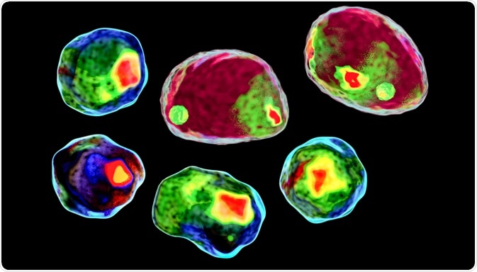
Cells produce various types of extracellular vesicles (EVs) that serve for short-range cell-to-cell communication in our bodies. As these EVs are usually less than one micrometer in size, the identification of different EV types can be a challenge.
 Image Credit: Kateryna Kon / Shutterstock.com
However, the latest flow cytometry (FC) methods are highly suitable for the analysis of different EV populations. Researchers from Italy have recently presented their optimized method for the FC analysis of extracellular vesicles from human mesenchymal stem cells (MSCs) in a protocol-publication, to share best practice with other researchers.
This protocol has been published in the journal Current Protocols in Stem Cell Biology.
The communication between cells in an organism is key to coordinate the cellular activity between different tissues. One well-known communication channel is represented by the endocrine system, where, for instance, hormones are released by specialized tissues to coordinate different organs in the body.
Besides the endocrine signaling, the paracrine system transmits information between cells over short ranges via extracellular vesicles. These EVs are biomembrane-enclosed vesicles that contain proteins and other biomolecules, and specific membrane proteins that reach to the outside can specifically interact with other cells.
There are at least three different subtypes of EVs, termed exosomes, microvesicles, and apoptotic bodies. While their different physiological functions are not fully understood, it has been observed that the different EVs differ in their sizes and densities.
The different structural features of the EV subtypes offer a way to separate them from each other, to conduct in-depth studies of their functions. Density-dependent separation of EVs by ultracentrifugation allows subsequent EV analysis by flow cytometry (FC), and an Italian research group led by Dr. Roberta Tasso (Genova) has recently shared their optimized EV purification method in a detailed protocol.
Separating extracellular vesicles based on density
Isolated mesenchymal stem cells in in vitro cultures secrete extracellular vesicles into their culture medium. EVs can be collected from the culture medium by density-gradient ultracentrifugation, where the different EV subtypes are separated according to their density.
Subsequently, the presence of EVs in the density-gradient solution can be determined by measuring the refractive index (RI) of the solution, as the EVs will cause a change of the RI.
Optimizing flow cytometry for EV analysis
Once EVs are isolated based on their density, flow cytometry can be used to characterize the individual EV subtype populations. However, as EVs are rather small (less than one micrometer in diameter), thorough calibration of the instrument is a prerequisite for an informative analysis.
The Italian researchers describe their best practice in FC calibration, and present examples of EV analyzes that demonstrate their ability to accurately determine the concentration and sizes of EV populations.
A key strength of common flow cytometry methods is that whole cells, or EVs can be labeled based on characteristic surface features, such as the presence of particular membrane proteins. Specific antibodies, conjugated to fluorescent molecules, can be used to mark EVs with a special surface feature. The scientists present examples for antibody-binding to three membrane proteins, measured on purified EVs.
The separation and FC analysis of different EV populations clearly allow more in-depth investigation of the paracrine functions of EVs in cell-to-cell signaling.
With the published methodology, which is fairly easy to use in most standard laboratories for cell-studies, best practice can be shared to drive research by making experimental research of different labs more comparable and reproducible.
Sources
Gorgun C et al. Isolation and flow cytometry characterization of extracellular-vesicle subpopulations derived from human mesenchymal stromal cells. Current Protocols in Stem Cell Biology 2019, 48, e76; DOI: 10.1002/cpsc.76.
Further Reading