Introduction
Blood Testing
Achieving Simultaneous Quantitative Phase and Absorption Measurements
Using Raman Spectroscopy to Provide Cellular Level Mapping of Blood Biochemistry
In Summary
References
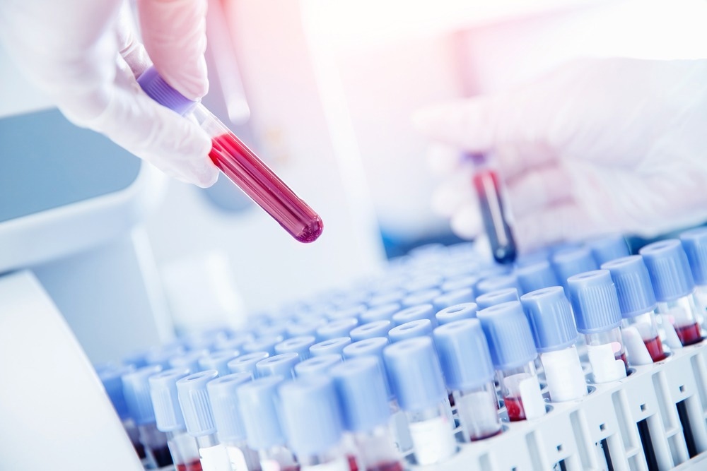
Blood testing is an essential medical process determining disease severity and organ dysfunction. This article will provide an overview and perspectives on mapping blood biochemistry at the cellular level.

Image Credit: Parilov/Shutterstock.com
Blood Testing
Blood testing detects the presence of harmful substances in a patient's blood that could cause severe medical complications during treatment, while taking medication, and pre-and-post surgical intervention. For instance, in chronic kidney disease, elevated calcium, bicarbonate, potassium, sodium, and uric acid concentrations can indicate the severity of renal dysfunction.
In diabetic patients, levels of blood glucose need to be constantly monitored to avoid severe health complications arising from their presence. The blood of patients with new-onset heart failure and chronic heart failure should be tested for electrolytes, hepatic enzymes, serum creatine, and other critical factors that indicate disease severity. Elevated lactate levels are associated with shock and poor prognosis in sepsis patients.
Over the years, clinical researchers have developed hundreds of blood testing procedures. Providing accurate and timely blood chemistry information helps clinicians perform earlier and more effective interventions that can improve patient outcomes and quality of life.
A routine blood test employed after surgery is a Chem 7 test, which tests for the presence of seven different substances in a patient's blood: carbon dioxide, creatinine, glucose, blood urea nitrogen, serum potassium, serum chloride, and serum sodium.
Blood chemistry analysis is conducted on a sample drawn from veins in fingers, arms, or earlobes, with some tests examining bone marrow cells. Using auto analyzers in laboratory tests allows clinicians to simultaneously perform several blood tests on the same sample.
Common blood test types include compatibility tests, sedimentation tests, complete blood counts, hemoglobin assays, and coagulation tests. Flow cytometry and impedance counters are commonly used in the clinical analysis of red blood cells. Techniques such as Raman imaging, FRET-based sensing, and gravitation separation have been developed in recent years.
Achieving Simultaneous Quantitative Phase and Absorption Measurements
Clinical translation has historically suffered from some major limitations at the cell level. One such limitation is the inability to simultaneously measure blood thickness and refractive index. Quantitative phase information is dependent on both these parameters. Imaging methods that use this information can improve the sensitivity and detail of blood cell analysis.
A study in 2011 provided beneficial research to overcome this limitation by decoupling the refractive index and thickness of red blood cells. The approach utilizes Spatial Light Interference Microscopy. The technique combined quantitative phase information with single absorption measurements, making it possible to analyze both at a cellular level.
Combined with morphological analysis, the technology enhances the analysis of blood cells, providing researchers with unprecedented detail and could enable new diagnostic capabilities for the detection and treatment of rare blood diseases.

Image Credit: Photoroyalty/Shutterstock.com
Using Raman Spectroscopy to Provide Cellular Level Mapping of Blood Biochemistry
Raman spectroscopy is a powerful analytical technique used in several industries to provide data on the structure of molecules and materials. It provides vibrational information on molecules to build a unique structural fingerprint to identify the target sample.
The technique offers an extremely sensitive diagnostic approach to determine a patient's complex cellular level blood biochemistry. Raman spectroscopy provides in vivo cellular biochemistry insights that provide information on blood samples' cellular environment.
Hemoglobin is a major component of mammalian blood, and analyzing this molecule's chemical structure is essential for monitoring patient blood health and providing information on several disorders.
One recent study published in 2022 demonstrated Raman spectroscopy's suitability for the analysis of blood heme color. Clinicians use the optical properties of blood to elucidate the health of patients. Blood's optical absorption behavior relates to the oxidation state of iron, which is a key component of hemoglobin. A fundamental understanding of the molecular dynamics of hemoglobin relies on the correlation between its electronic distribution and physical structure.
Raman spectroscopy is sensitive to heme states, and this characteristic of the technique allows researchers to probe distributions of different forms of hemoglobin and their dependence on cellular physiology.
Combining Raman spectroscopy with fluorescence microscopy helps to correlate hemoglobin redistribution with ionic signaling and cell dynamics. This technique can aid the development of new hematology protocols by evaluating the response of cell structural reactivity to biochemical and biophysical interventions.
In Summary
Monitoring and assessing blood biochemistry is central to disease diagnosis and the evaluation of treatments and medical interventions. Over the years, several powerful analytical techniques and blood assays have been developed to improve blood biochemistry analysis and patient prognosis. This article has provided a brief overview of current perspectives on this crucial area of biomedical research.
Read Next: Using Flow Cytometry to Analyze Peripheral Blood Cells
References:
Further Reading