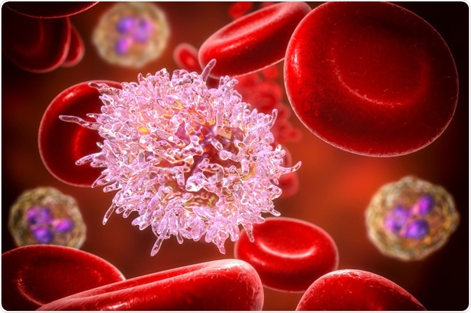
Anaplasia describes cells which are undifferentiated, or a poorly differentiated. This means that they lose characteristics associated with a certain tissue (which is made of cells).
 Image Credit: Kateryna Kon / Shutterstock
Typically, anaplastic cells have hyperchromatic nuclei, prominent nucleoli, a nucleus:cytoplasm size ratio which is close to 1:1, and they show increased mitotic cell division.
The nucleus is where the chromosomes sit within a eukaryotic cell. The chromosomes are the compressed version of chromatin, and is formed of DNA and proteins.
Hyperchromatism, in this instance, refers to the development of excess chromatin, and shown as increased staining in the nucleus.
This means that anaplastic cells and tissues are visually different to non-anaplastic cells and tissues because of the lack of differentiation, and the cells can be of various sizes including giant cells.
The nucleus of anaplastic cells may stain darker and are often larger than those of non-anaplastic cells due to hyperchromatism. Cellular orientation and organization can also be altered in anaplastic tissues, and evidence of mitosis may be seen more frequently in anaplastic cells.
Causes of anaplasia in thyroid cancer
Anaplastic thyroid cancer occurs less frequently than other thyroid cancers but is associated with the majority of deaths from thyroid cancers. To see what genetic factors are involved in anaplastic thyroid cancer, mouse models were used.
Commonly, two signaling pathways are de-regulated in anaplastic thyroid cancer: the PI3K signaling pathway and the mitogen-activated protein kinase (MAPK) signaling pathway. Therefore, it can be assumed that these pathways play a role in the development of anaplastic thyroid cancer.
The PI3K pathway
The PI3K pathway is crucial for the uptake of glucose into cells, cell growth and proliferation, cell adhesion, cell motility and survival. Activation of receptors by extracellular ligands activates PI3K, which causes the formation of PIP3 by phosphorylation.
PIP3 then localizes AKT to the membrane, where PDK1 activates AKT. AKT is the effector of this pathway, and controls many biological responses. PIP3CA is mutated in 15 – 25% of anaplastic thyroid cancers, and it is also amplified in 40% of anaplastic thyroid cancers.
The MAPK pathway
The MAPK signaling pathway is crucial for processes such as cell growth and proliferation, differentiation, migration and apoptosis. 70% of thyroid cancers have been shown to have changes in the MAPK signaling pathway.
RAS proteins are a component of the MAPK signaling pathway, acting as a switch that can be activated when the receptors on the cell membrane are triggered.
Mutations in RAS are seen in anaplastic thyroid cancer, and varies in prevalence from 8 - 60%. RAF proteins, which RAS act on, are also mutated in anaplastic thyroid cancer; the RAF protein B-RAF is the most commonly mutated, and is seen in 25 – 44% of anaplastic thyroid cancers.
Causes of anaplasia in medulloblastoma
Medulloblastoma is a type of brain tumor which is the most common “central nervous system embryonal tumor”, accounting for 15 – 20% of childhood brain tumors. Anaplasia in medulloblastoma has been correlated with a negative prognosis. Another negative prognosis indicator was the increase in the expression of c-myc gene.
As this was only observation, and no link had been established between anaplasia and increased c-myc expression in medulloblastoma, Stearns and co. sought to see if there was indeed a link. The authors used two types of medulloblastoma cells, the cell lines DAOY and UW228.
These were chosen as they did not have c-myc over-expression. The c-myc gene was purposefully stably overexpressed in both cell lines, and found that these cells grew faster than cells which were not over-expressing c-myc.
These cells were then injected into mice to form tumors, and it was found that DAOY cells over-expressing c-myc gave rise to tumors which were 75% larger compared to the tumors derived from DAOY cells that were not over-expressing c-myc.
UW228 cells that were not over-expressing c-myc did not form tumors. Interestingly, when these tumors from mice were analyzed, the tumors from cells over-expressing c-myc showed sever anaplasia, including changes such as larger nuclei. Therefore, c-myc expression seems to be linked to anaplasia in medulloblastoma.
Further Reading